Vision is a precious sense that shapes daily life in countless ways. When the eyes are healthy, it's easy to take that clarity for granted. However, even minor eye problems can interfere with essential tasks like reading or driving and more serious conditions may threaten the gift of sight altogether. That's where ophthalmology comes in.

What Is Ophthalmology?
Ophthalmology is the medical specialty focused on diagnosing, treating and preventing disorders of the eye. An ophthalmologist is a physician who has completed medical school plus advanced training in eye-related care. This training covers everything from routine vision checks to complex surgeries. For instance, an ophthalmologist can prescribe corrective lenses for nearsightedness, remove cataracts that cloud vision or manage systemic conditions like diabetes when they affect the eyes. Their broad scope sets them apart from other eye care professionals: they are the only providers with full medical and surgical authority over a range of eye conditions, including advanced diseases such as macular degeneration, diabetic retinopathy and glaucoma. Although some eye problems cause immediate symptoms, many progress silently. For that reason, ophthalmologists collaborate closely with other healthcare specialists. Patients with high blood pressure or diabetes often need coordinated care because these conditions can damage the small blood vessels that nourish the retina. By detecting issues early and intervening appropriately, ophthalmologists preserve vision and help patients keep independence, whether through medication, lifestyle recommendations or advanced procedures.
What Are the Major Eye Diseases in Ophthalmology?
Ophthalmology addresses a wide range of conditions that can affect the eye's structures, including the cornea, lens, retina and optic nerve. Some of the most prevalent causes of vision loss have been well studied and typically respond to timely treatment.
- Cataract
A cataract develops when the eye's natural lens becomes cloudy or opaque, leading to blurry vision and duller colors. These changes often progress gradually, especially in older adults. Because the lens is crucial for focusing light onto the retina, any clouding will affect a person's ability to read, drive or recognize faces. Although cataracts are the most common cause of reversible blindness worldwide, modern cataract surgery can remove the cloudy lens and replace it with a clear artificial lens (intraocular lens or IOL). This operation boasts high success rates, allowing the vast majority of patients to enjoy sharper, more vibrant vision.
- Glaucoma
Glaucoma is a group of diseases defined by damage to the optic nerve, often in the presence of increased intraocular pressure. This condition tends to progress slowly and quietly, with early stages typically free of any symptoms. Over time, glaucoma can erode side vision and eventually central vision. Because it is a leading cause of irreversible blindness worldwide, early detection is critical. Regular screening (especially if there's a family history or other risk factors) helps find patients who may receive help from eye drops, laser procedures or surgery to lower eye pressure and preserve the optic nerve.
- Age-Related Macular Degeneration (AMD)
AMD damages the macula, the central portion of the retina responsible for detailed sight. People with AMD often retain their peripheral vision but experience difficulty recognizing faces, reading small print or driving. There are two types: dry AMD, characterized by gradual thinning of the macula and the presence of small deposits called drusen and wet AMD, which involves abnormal blood vessel growth beneath the retina. Wet AMD can lead to rapid, severe vision changes if not addressed. While there is no complete cure for advanced forms, treatments such as anti-VEGF injections can slow the progression of wet AMD and help patients retain more central vision.
- Diabetic Retinopathy
Diabetes can harm the blood vessels that supply the retina, causing a spectrum of complications known as diabetic retinopathy. Elevated blood sugar weakens vessel walls, making them prone to leaking or growing abnormally. Early stages may cause few or no symptoms, but as the disease progresses, patients might notice blurred vision, dark spots or sudden loss of sight. Tight control of blood sugar and blood pressure can delay onset or slow progression. When retinopathy becomes advanced, treatments such as laser photocoagulation, anti-VEGF injections or vitrectomy surgery help prevent more severe vision loss. Annual eye exams are recommended for those with diabetes, since early intervention is key to preserving sight.
Which Diagnostic Tools Are Commonly Used in Ophthalmology?
Ophthalmology relies on specialized devices and imaging methods that reveal detailed information about the eye's delicate structures. Early detection of disease is often the difference between saving and losing sight, so precise diagnostic tools play a central role.
- Optical Coherence Tomography (OCT)
OCT uses light waves to capture cross-sectional images of the retina and optic nerve with microscopic detail. By scanning the layers of the retina, ophthalmologists can detect swelling, fluid accumulation or thinning that shows disease progression. This imaging is crucial for checking wet AMD, diabetic macular edema and glaucoma. Because OCT is noninvasive and gives clear, detailed views, it has become a standard for diagnosing and tracking many retinal and optic nerve conditions over time.
- Fundus Photography
Fundus photography provides color images of the retina, optic disc and macula. These photographs offer a window into changes at the back of the eye that might signal diabetic retinopathy, AMD or glaucoma. Patients may see images of their own eye structures, which can be helpful in understanding their condition. Additionally, these images serve as documentation for future comparisons, allowing ophthalmologists to see if treatments are collaborating or if a disease has progressed.
- Visual Field Testing (Perimetry)
Visual field tests measure the sensitivity of peripheral vision. While people are typically aware of what they see straight ahead, side vision can deteriorate unnoticed. A key application of perimetry is checking glaucoma, which slowly erodes peripheral vision. The test involves watching for lights flashed in different areas of the field of view. Areas where the patient consistently misses lights may represent nerve fiber loss. Neurological conditions that affect the visual pathways, such as certain brain tumors or strokes, can also be found using this method.
- Tonometry and Other Tests
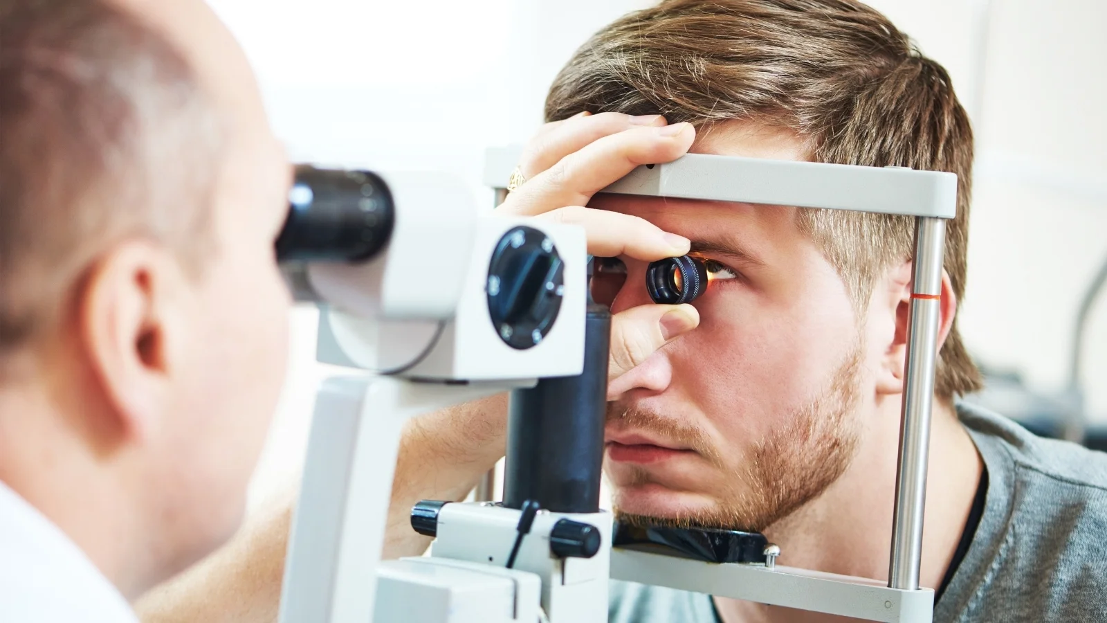
Measuring intraocular pressure (tonometry) is foundational in glaucoma screening. Ultrasound biomicroscopy can be used when the view into the eye is blocked by a dense cataract or hemorrhage. Corneal topography is particularly helpful in mapping the curvature of the cornea for refractive surgery planning or diagnosing corneal diseases like keratoconus. Comprehensive exams also typically include a slit-lamp evaluation of the cornea, iris and lens under magnification. By integrating all these data points, ophthalmologists gain a comprehensive view of ocular health and can tailor treatment plans accordingly.
What Are the Most Common Treatments and Surgeries in Ophthalmology?
Management of eye diseases often involves a combination of medications, laser therapy and surgical interventions. Ophthalmologists customize treatment based on factors like disease severity, patient lifestyle and overall health status.
- Cataract Surgery
When cataracts interfere with daily life, surgical removal is the only definitive remedy. During this brief outpatient procedure, the cloudy lens is fragmented (often using ultrasound technology called phacoemulsification), removed and replaced by an artificial IOL. Patients usually see a significant improvement in clarity, contrast and color perception. Advances in technology also allow for laser-assisted cataract surgery, which can enhance precision during certain steps. Furthermore, modern IOLs may correct astigmatism or reduce dependence on reading glasses.
- LASIK and Other Refractive Procedures
LASIK aims to reduce or cut the need for glasses or contact lenses by reshaping the cornea with a laser. After creating a thin flap, the surgeon sculpts the corneal tissue to correct nearsightedness, farsightedness or astigmatism. PRK (photorefractive keratectomy) and SMILE (Small Incision Lenticule Extraction) are alternative procedures that achieve similar results through different techniques. For many, these outpatient surgeries are life-changing, offering improved visual acuity within days and minimal downtime. Because laser-assisted corrections have become routine, there is a strong track record of safety and efficacy dating back decades.
- Intravitreal Injections
A growing number of retinal conditions receive help from medication delivered directly into the vitreous cavity. Anti-VEGF drugs, such as bevacizumab, ranibizumab or aflibercept, collaborate by blocking a protein that promotes abnormal blood vessel growth and leakage. These injections can stabilize and sometimes improve vision in conditions like wet AMD, diabetic macular edema or retinal vein occlusions. While the idea of a shot into the eye may sound intimidating, the procedure is usually brief and tolerable under local anesthesia. In most cases, repeated injections over months or years are needed to keep receives help.
- Glaucoma Surgeries
When eye drops or laser therapy do not sufficiently control intraocular pressure, surgical choices come into play. Traditional surgeries, like trabeculectomy or tube shunt implants, create new pathways for fluid drainage to lower pressure. However, minimally invasive glaucoma surgeries (MIGS) have gained popularity for their lower risk and faster recovery time. These procedures use tiny stents, shunts or micro-incisions to enhance fluid outflow. While MIGS may not reduce pressure to the same extent as traditional methods, they offer a safer profile and can be combined with cataract surgery.
What Recent Innovations Are Transforming Ophthalmology?
Ophthalmology has been at the forefront of medical innovation, embracing advanced imaging, novel therapeutics and refined surgical procedures. These advancements often lead to earlier intervention, improved safety and better outcomes for patients.
- Gene Therapy for Inherited Retinal Diseases
A groundbreaking moment in ophthalmology came when a gene therapy was approved for a specific type of childhood blindness caused by mutations in the RPE65 gene. By injecting a healthy copy of the gene directly into retinal cells, the therapy enables production of a crucial enzyme, allowing some patients to recover functional vision. This success has sparked numerous clinical trials targeting other inherited retinal disorders. Researchers are also investigating stem cell treatments that might replace lost retinal cells and further extend therapeutic possibilities for conditions once deemed untreatable.
- Minimally Invasive Glaucoma Surgery (MIGS)
MIGS represents a paradigm shift for glaucoma treatment. Microscopic devices, such as the iStent or Xen gel stent, offer earlier and safer surgical intervention by creating or enhancing fluid drainage in ways that disrupt less tissue than older surgeries. Even though these implants may produce more modest pressure reductions than more extensive procedures, they significantly lower the risk of complications. As a result, MIGS is particularly suited for mild to moderate glaucoma, often performed at the same time as cataract surgery for convenience. Ongoing refinements in device design promise to improve pressure control further and expand access to safer glaucoma management.
- Artificial Intelligence (AI)
Machine learning has demonstrated remarkable potential in analyzing retinal images. Certain AI algorithms can detect the earliest signs of diabetic retinopathy in fundus photographs with accuracy comparable to seasoned clinicians. Some systems are approved for use in settings without an ophthalmologist on-site, enabling primary care clinics to offer timely screening and referral for advanced care. AI also holds promise for predicting which patients are at higher risk for rapid disease progression, guiding more personalized treatment plans. In glaucoma management, AI could assist with large-scale analysis of visual field tests and OCT images to predict nerve damage. Rather than replace human skill, these tools serve as a valuable complement that can free up specialists for more nuanced clinical decision-making.
- Advanced Imaging and Surgical Techniques
OCT Angiography (OCT-A) allows the visualization of retinal vessels without injecting dye, providing a clearer, noninvasive look at blood flow beneath the macula. This technique can detect very subtle vascular changes in wet AMD or diabetic retinopathy. Femtosecond lasers are increasingly used for precise corneal incisions in cataract surgery and refractive procedures like LASIK, boosting accuracy and reducing healing time. Efforts to develop retinal implants or "bionic eyes," aim to restore basic visual perception for those with advanced retinal degeneration. Though these implants are still in development, they offer hope for patients who have been blind for many years.
Why Are Regular Eye Exams Essential in Ophthalmology?
Routine eye exams allow for early detection and intervention, which is especially important because many threatening conditions progress quietly. Glaucoma is known as the "silent thief of sight" because it gradually erodes peripheral vision before patients notice any loss. Diabetic retinopathy and early-stage macular degeneration can also stay asymptomatic for years. A comprehensive eye exam may involve dilating the pupils, checking eye pressure and assessing retinal health.
- For adults without known risk factors, national guidelines often recommend eye exams every few years in early adulthood, with increasing frequency as people get older. After age 40, the risk of cataract, glaucoma and AMD starts to rise, so more regular checkups become prudent. By age 65, it's generally recommended to have annual or biennial exams to catch treatable issues early.
- People with diabetes should have a dilated eye exam at least once a year because diabetic retinopathy can advance rapidly if blood sugar is poorly controlled. Family history of glaucoma or macular degeneration may also call for earlier and more frequent evaluations. Children receive help from early vision screenings, typically performed during routine pediatric visits. Detecting issues like a lazy eye or refractive errors in childhood can improve outcomes when treated promptly.
In many cases, taking action before symptoms arise can preserve years of healthy vision. Eye examinations can also reveal clues about general health; for instance, changes in the retinal blood vessels might show poorly controlled hypertension or diabetes. By emphasizing prevention and vigilant checking, ophthalmology helps patients keep a lifetime of good eyesight.


